

Nanomedicine can potentially result in more effective drugs with fewer side effects. But the road to that end is lined with challenges.
Text & Photo: Nicklas Hägen
Within nanomedicine size is everything. This field is all about working with synthetic particles which are so small that the characteristics of the material are changed. The utilisation of these characteristics in a controlled way is called nanotechnology, and nanomedicine consequently means ‘the medical application of nanotechnology’.
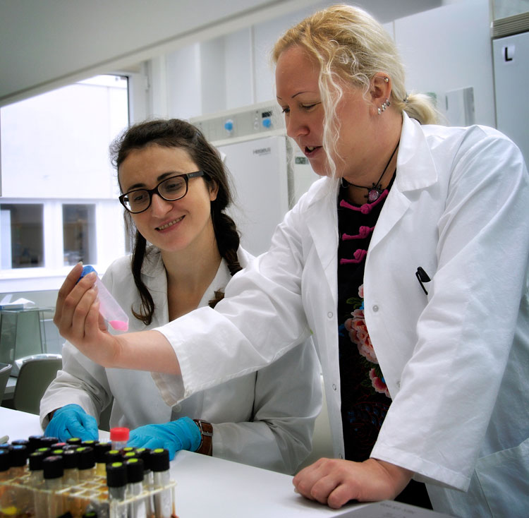
“The characteristics that change can be electric, magnetic or chemical, but they always pertain to size,” says Jessica Rosenholm, professor in pharmaceutical development at Åbo Akademi University and a specialist in nanomedicine.
“When the particles become small enough, they acquire characteristics that the bulk material does not have. As the size of gold particles, for example, shrinks to under a hundred nanometres their colour starts to change because their capacity for light refraction changes. The optical features of nanomaterial can be utilised in, for instance, contrast agents for medical examinations.”
Nanoparticles are smaller than can be discerned with the naked eye, but they are counted as material. Roughly speaking this means that they are considerably larger than single molecules – or amalgamations of molecules, which in turn, to put it simply, are formed of bonds between atoms of various chemical elements.
While the particles are larger than both molecules and atoms, they are so small that they are not influenced by gravity to any significant degree. And compared to body cells, the nanoparticles are tiny.
One reason as to why its chemical characteristics change when a particle becomes sufficiently small is that a relatively large proportion of the atoms are situated on the surface of the particle instead of being bound to each other inside it. With many atoms on the surface, a particle is very reactive (to such an extent that it might even cause so-called nanotoxicity, i.e. health risks). In very small particles most atoms can, in fact, be situated on its surface.
“We are interested in nanoparticles because they are approximately as small as the body’s own biomolecules. This means that they can circulate around the body. The body might not recognise nanoparticles as foreign material, as they are small enough and they can be designed in such a way that the immune system doesn’t recognise them,” says Rosenholm.
“Nanoparticles can be accumulated in tissue and absorbed by cells. That is why we’re interested in using them as drug carriers. We can fill them with pharmaceuticals – for example cytostatics that are aimed only at the tumour and not at healthy cells, as this leads to side effects. If we’re able to send the drug exclusively to the cancer cell, only that cell is affected by the cytotoxin.”
Not a utopian dream
Nanopharmaceuticals are not a new invention and not a utopian dream, either. Research has been carried out since the 1980s and the first nanodrug was launched on the market in 1995. It was a cancer drug based on liposomes, small fat-like bubbles made of the same material as the cell membrane.
As technology has developed and improved, researchers are now able to start realising the potential of nanomedicine. Nanopharmaceuticals are being developed for various therapeutic purposes and there are already, for example, vaccines in nanoform and nanodrugs for diabetes, cardiovascular diseases and neurodegenerative diseases at various phases of development. The most common, however, are cancer drugs.
“Nanomedicine gives many, many so far unexploited possibilities. Pharmaceutical development is a slow process. It takes about 15 years from the date of a new discovery to a new product being launched on the market. After the boom of nanotechnology a number of new and different nanomaterials have emerged which have not yet reached the market. But they’re on their way,” says Rosenholm.
“The pharmaceuticals already on the market, that is, the first generation of nanodrugs, are not entirely selective. They do transport more effectively around the body than if the medical substance was given on its own without the liposome as a carrier system, but they could be more selective and more effective, and include more functions, for example abilities such as actively seeking their target, visualising and circumventing drug resistance.”
Carrier systems
The possibility of targeting the delivery of traditional drugs in the body is limited. After a pharmaceutical substance is dissolved into the body from, for example, a pill, its subsequent fate is dependent on the chemical and physical characteristics of the drug molecule. The possibilities of prior manipulation of the drug molecule in order to direct it towards the destination where it is intended to have an impact are small, since every operation changes the molecule and thus also its medical effect.
Nanopharmaceuticals provide an opportunity to direct the movements and absorption of the drug in the body. There are, nevertheless, several difficulties involved in doing this. First of all, the immune system must be ‘cheated’ into not recognising the drug-carrying particle as something foreign to the body, but rather allowing it to circulate for long enough to find its way to the place where it is intended to have an effect. Secondly, the substance must be efficiently accumulated and released in the appropriate place in the body. And thirdly the destination of the pharmaceutical molecule is often a protein in the cell, and therefore the aim is that the cell shall absorb the drug, which is to find its correct place in the cell.
In order for this to succeed a so-called ‘carrier system’ is created, consisting of particles with the appropriate chemical and physical characteristics for all three aspects to function. These particles are then ‘loaded’ with the molecules of the pharmaceutical substance – there might be room for millions of them in one nanoparticle.
Chemical design provides many opportunities for giving the carrier particle the desired characteristics.
“We try to make the carrier system behave in the way we want it to. After that we can basically load the particle with any drug molecule without affecting the carrier system. The system takes the drug molecule to its set destination and releases it there,” says Rosenholm.
“The exact way of doing this is a question of nanoparticle design, which is what we focus on in our daily work at the lab. It involves playing with chemistry,” Rosenholm explains.
One way of creating the particles is to start concretely with a large block of the material, which is ground into small particles, but Rosenholm’s research group starts from the opposite direction, building their particles molecule by molecule. The carrier systems they create usually consist of porous ceramic silicon dioxide particles.
“We begin with solutions that we mix together. The components included in the solution are molecular and separate from each other, but when they are mixed, they start to accumulate and react with each other, becoming linked to larger and larger elements, particles. By playing with temperature, solvents and pH values we can see to it that they don’t grow too large,” says Rosenholm.
“Our silicon dioxide particles are porous. When the particles are completed, we put the drug substance into the pores. We prepare a solution of the drug, add the particles and let the two merge overnight. It’s crucial that the surface chemistry of the particle pore walls is such that it attracts the drug substance. By using a ‘poor’ solvent the drug molecules will rather be absorbed by the particles than stay in the solvent. Then we can remove the particles from the solution using a centrifuge, vacuum dry them and – voilà! – we have a completed nanopharmaceutical.
Tissues and contrast agents
In addition to the development of pharmaceuticals, nanomedicine comprises two other research areas. One is diagnostics, where nanotechnology is used to create contrast agents. The other is regenerative medicine, where various tissues or organs are created in a laboratory environment – so-called tissue reconstruction. Professor Jessica Rosenholm is also involved in research projects at Åbo Akademi University which are connected to both of these areas.
The aim of tissue reconstruction is 3D printing of organs or tissue to be used, for example for prostheses or wound healing. These can be implanted with cells and nanodrugs in order for them to grow into the body.
Niklas Sandler, professor in pharmaceutics at Åbo Akademi, studies printable pharmaceuticals. In cooperation with him, Rosenholm has already been able to print 3D model prostheses made of synthetic polymers. The aim, within a collaborative project with Stefan Willför, professor in wood and paper chemistry at Åbo Akademi, is to expand materials to include natural, biocompatible polymers such as cellulose and hemicellulose. For further applications cooperation is carried out with Cecilia Sahlgren, group leader and senior scientist at the Turku Centre for Biotechnology and one of a group of experts on tissue regeneration at the Eindhoven University of Technology in the Netherlands.
“Nanoparticles are mixed with biodegradable polymers and the pulp with the particles is then printed. The particles can contain pharmaceutical substances which regulate the regeneration of the tissue around the prosthesis by making sure that the cells differentiate in the appropriate direction and develop into the cells we want them to be. Regenerative medicine can be about regenerating any organ or tissue, but the cell differentiation must be regulated so that the right cells are created for the right organ,” Rosenholm explains.
Rosenholm’s research group also makes nanodrugs which can both be visualised and carry pharmaceutical substances. As these can be studied in both cells and animal models, they give research a new dimension as the drug can be traced and its effect can be followed up.
At the same time contrast agents are developed that can be monitored using several different methods.
“The different imaging techniques supplement each other as to resolution and sensitivity, that is, they are crucial for the level that can be looked at. This makes it possible to study the same sample at various levels: with optical methods at the cellular level and with, for example, a microscope or magnetic resonance imaging at organism level.”
Is there a risk of getting different results if using different samples for different methods?
“Yes, there is. You can never get an exact copy of biological systems.”